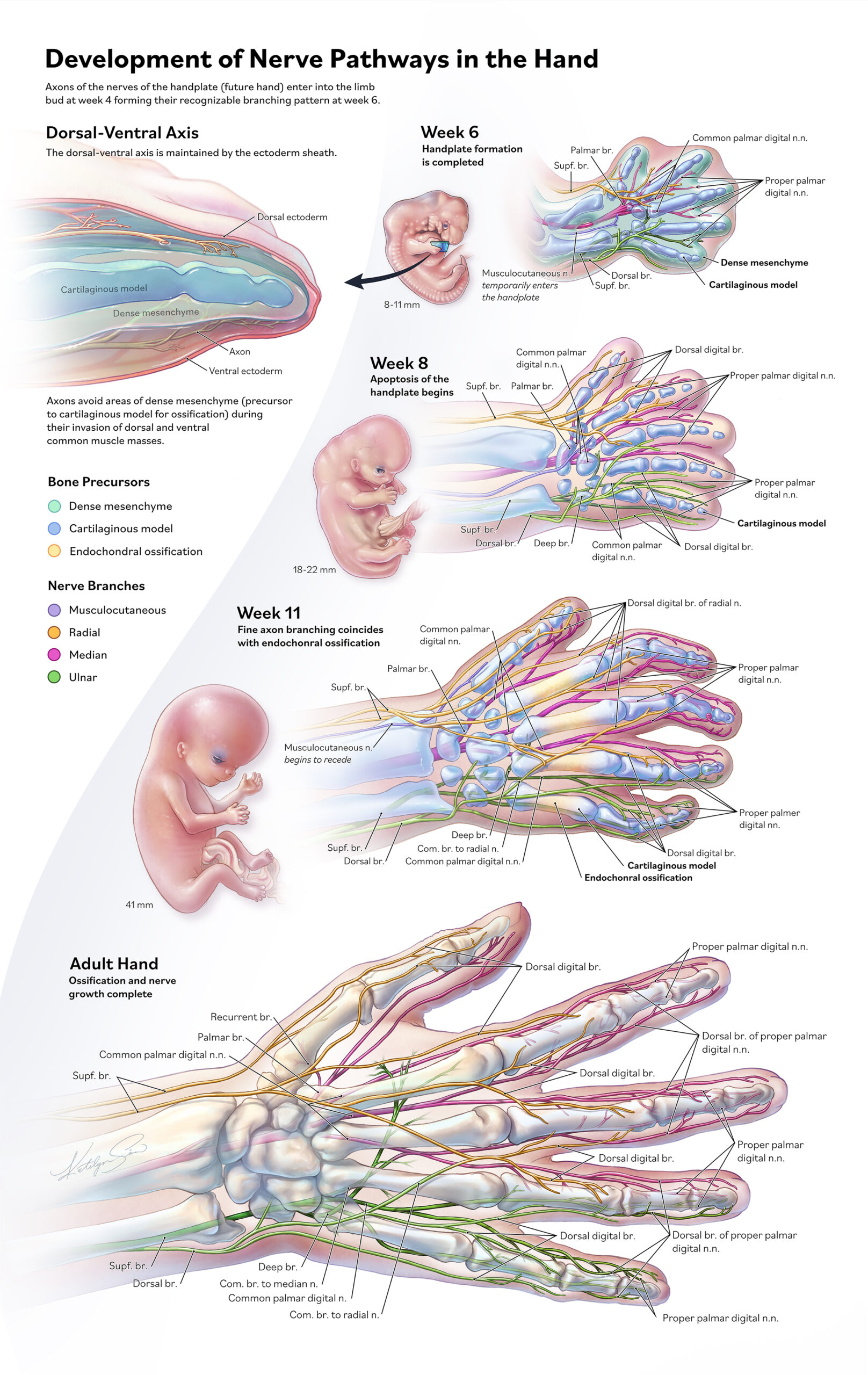
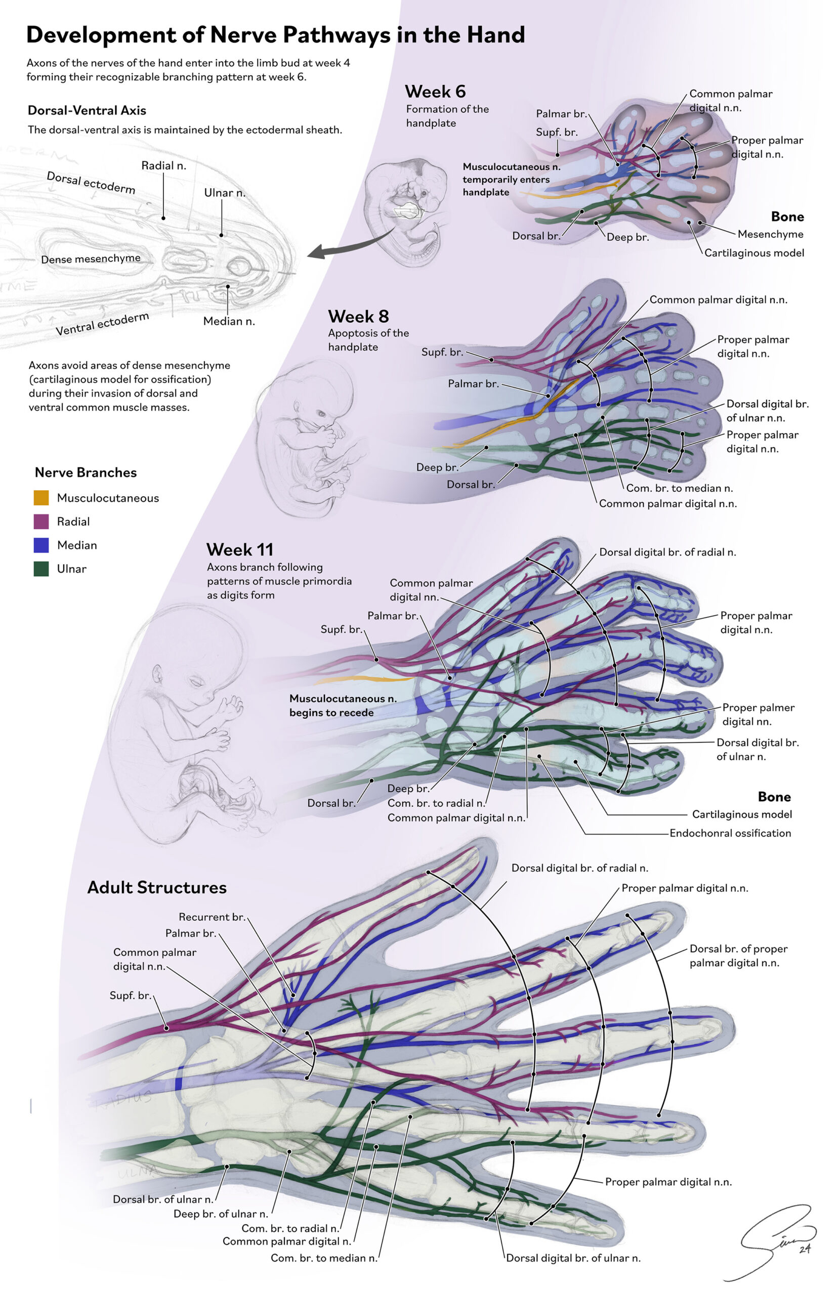
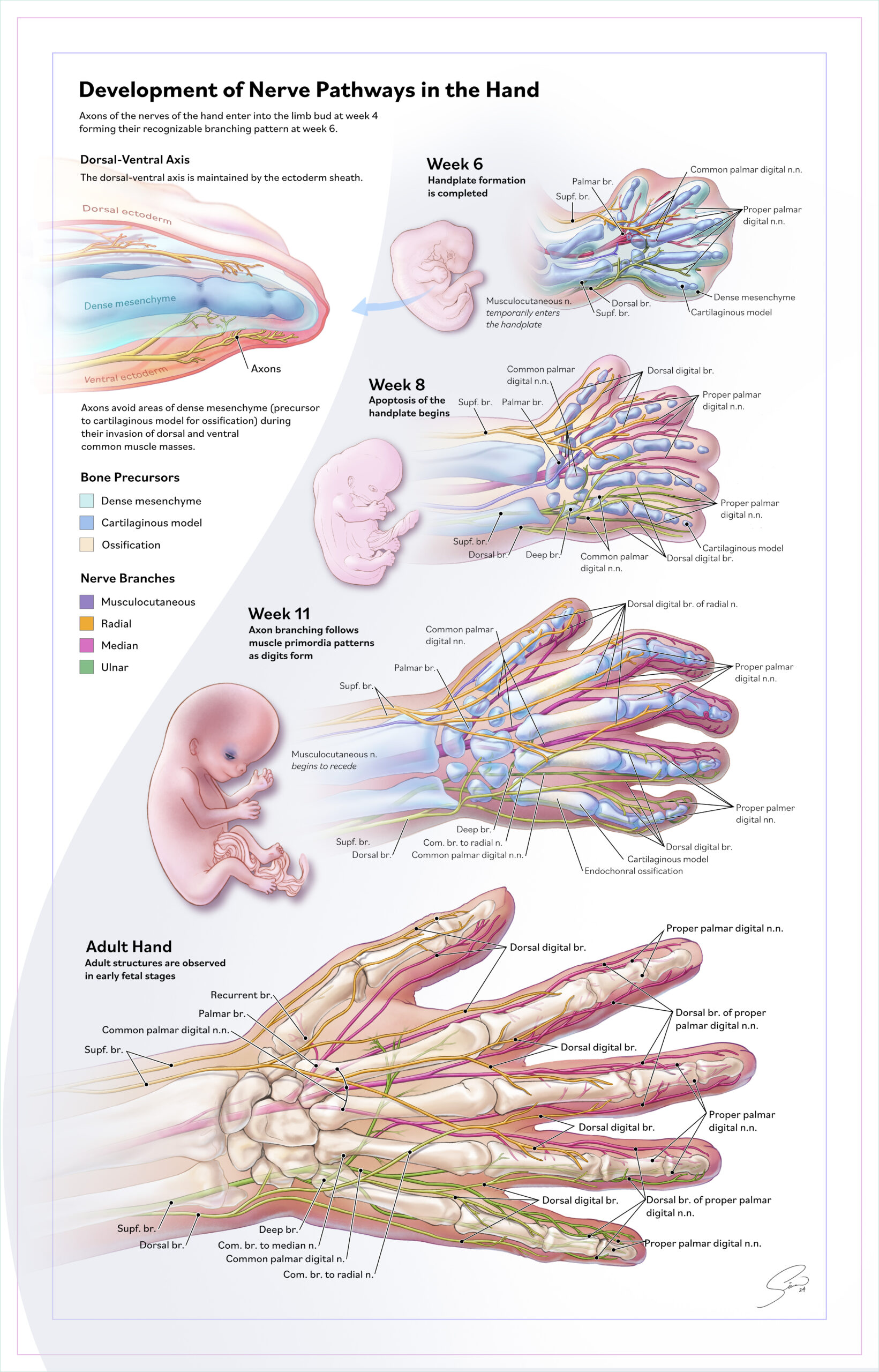
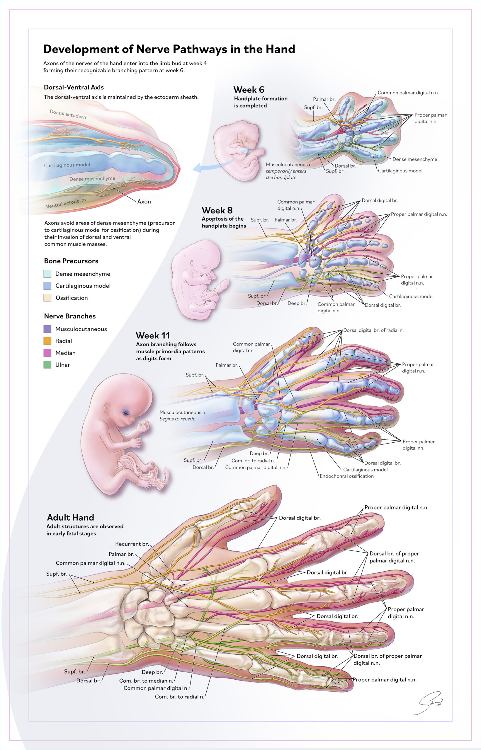
What I found most fascinating was how nerves establish pathways based on the density of undifferentiated stem cell tissue (mesenchyme), growing into anticipated routes while avoiding areas of dense mesenchyme that later ossify into bone. This early guidance sets the stage for the complex integration of nerves, bones, and cartilage in the mature hand. To make these relationships clear for teaching, I used color coding to differentiate nerve branches and bone precursors, while also orienting the work along the dorsal–ventral axis.
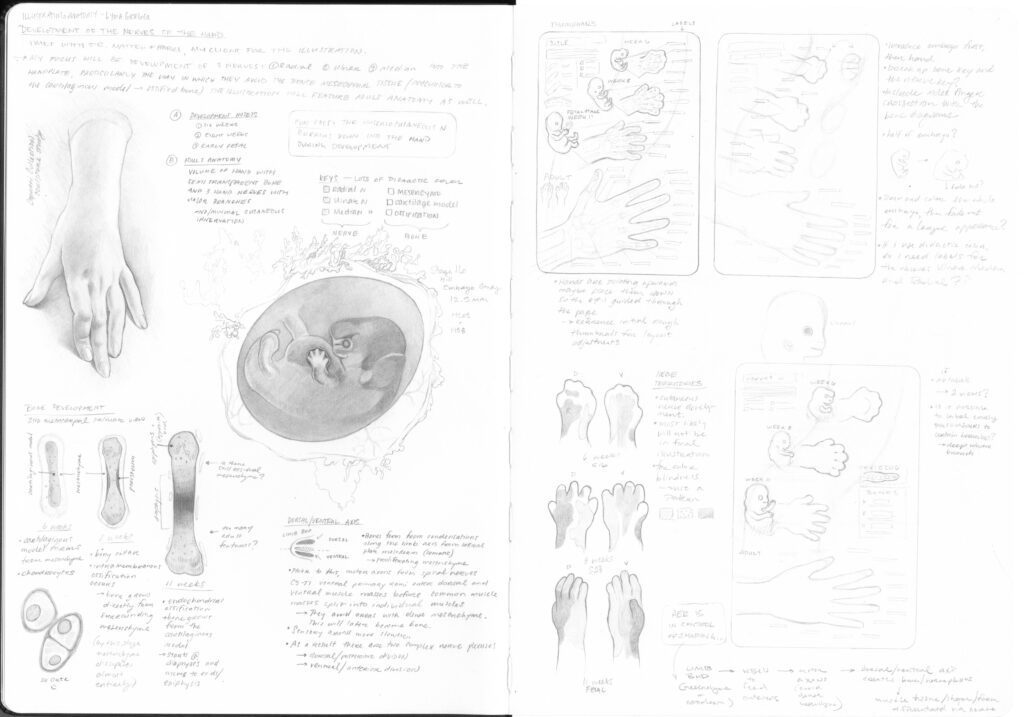
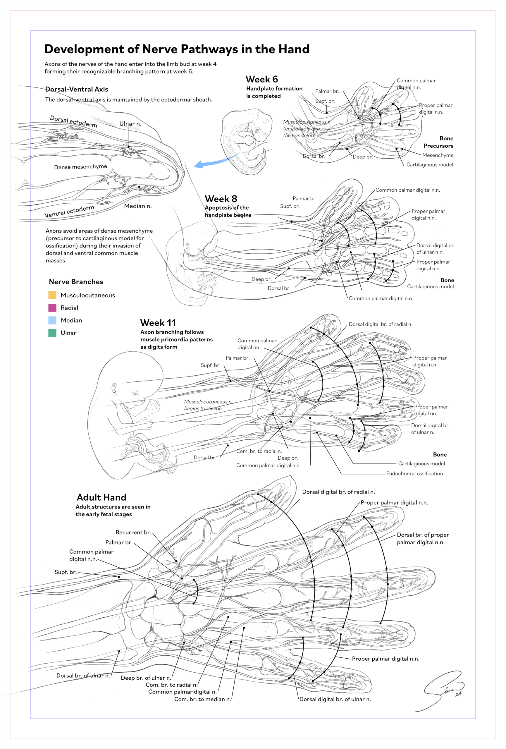
When drawing thumbnails, I knew I would have to sacrifice having both a dorsal and ventral view of each hand, so I focused on creating an overlapping network of dorsal and ventral nerves using depth, shadow, and the developing bone as a margin for anterior and posterior. We decided that having 3 stages would best tell our visual story:
Week 6 (Carnegie Stage 16) – the hand plate begins to web
Weeks 7–8 (Carnegie Stages 20–23) – apoptosis sculpts the digits
Early Fetal Stage (11 weeks) – more refined branching of nerves with early skeletal structures